Diffraction project datasets GS14013A_3g6i
Project details
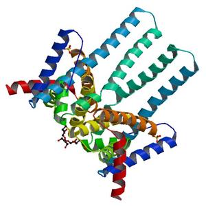
| Title |
Crystal structure of an outer membrane protein, part of a putative carbohydrate binding complex (bt_1022) from bacteroides thetaiotaomicron vpi-5482 at 1.93 A resolution |
| Authors |
Joint Center for Structural Genomics (JCSG) |
| Bioentity |
None |
| R / Rfree |
0.20 / 0.22 |
| Unit cell edges [Å] |
75.03 x
75.03 x
127.29
|
| Unit cell angles [°] |
90.0,
90.0,
90.0
|
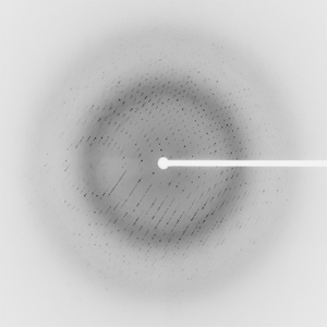
Dataset 106933_1_E1_###.mccd details
| Number of frames |
90 (1 - 90) |
| Distance [mm] |
250.0 |
| Oscillation width [°] |
1.00 |
| Phi [°] |
237.0 |
| Wavelength [Å] |
0.97964 |
| Experiment Date |
2009-01-17 |
| Equipment |
BL9-2
at SSRL (Stanford Synchrotron Radiation Laboratory)
|
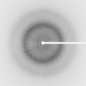
Dataset 106933_1_E2_###.mccd details
| Number of frames |
90 (1 - 90) |
| Distance [mm] |
250.0 |
| Oscillation width [°] |
1.00 |
| Phi [°] |
237.0 |
| Wavelength [Å] |
0.91837 |
| Experiment Date |
2009-01-17 |
| Equipment |
BL9-2
at SSRL (Stanford Synchrotron Radiation Laboratory)
|
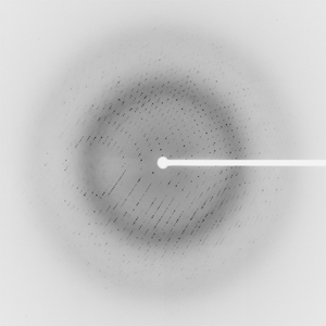
Dataset 106933_2_###.mccd details
| Number of frames |
90 (1 - 90) |
| Distance [mm] |
250.0 |
| Oscillation width [°] |
1.00 |
| Phi [°] |
237.0 |
| Wavelength [Å] |
0.97949 |
| Experiment Date |
2009-01-17 |
| Equipment |
BL9-2
at SSRL (Stanford Synchrotron Radiation Laboratory)
|
Dataset 106933_3_###.mccd details
| Number of frames |
90 (1 - 90) |
| Distance [mm] |
350.0 |
| Oscillation width [°] |
1.00 |
| Phi [°] |
237.0 |
| Wavelength [Å] |
0.91837 |
| Experiment Date |
2009-01-17 |
| Equipment |
BL9-2
at SSRL (Stanford Synchrotron Radiation Laboratory)
|