Diffraction project datasets GS13859A_3d02
Project details
| Title |
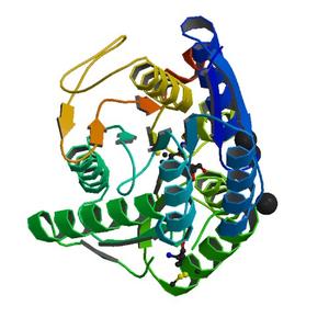
Crystal structure of periplasmic sugar-binding protein (YP_001338366.1) from Klebsiella pneumoniae subsp. pneumoniae MGH 78578 at 1.30 A resolution |
| Authors |
Joint Center for Structural Genomics (JCSG) |
| Bioentity |
None |
| R / Rfree |
0.16 / 0.17 |
| Unit cell edges [Å] |
66.24 x
76.96 x
113.98
|
| Unit cell angles [°] |
90.0,
90.0,
90.0
|
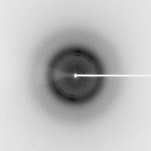
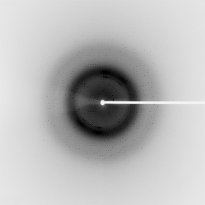
Dataset 81258_1_E1_###.mccd details
| Number of frames |
400 (1 - 400) |
| Distance [mm] |
190.0 |
| Oscillation width [°] |
0.25 |
| Phi [°] |
220.0 |
| Wavelength [Å] |
0.97932 |
| Experiment Date |
2008-03-16 |
| Equipment |
BL9-2
at SSRL (Stanford Synchrotron Radiation Laboratory)
|
Dataset 81258_1_E2_###.mccd details
| Number of frames |
400 (1 - 400) |
| Distance [mm] |
190.0 |
| Oscillation width [°] |
0.25 |
| Phi [°] |
220.0 |
| Wavelength [Å] |
0.91162 |
| Experiment Date |
2008-03-16 |
| Equipment |
BL9-2
at SSRL (Stanford Synchrotron Radiation Laboratory)
|
Dataset 81258_2_###.mccd details
| Number of frames |
400 (1 - 400) |
| Distance [mm] |
190.0 |
| Oscillation width [°] |
0.25 |
| Phi [°] |
220.0 |
| Wavelength [Å] |
0.97918 |
| Experiment Date |
2008-03-16 |
| Equipment |
BL9-2
at SSRL (Stanford Synchrotron Radiation Laboratory)
|