Diffraction project datasets GS13645A_3h3l
Project details
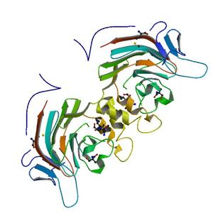
| Title |
Crystal structure of PUTATIVE SUGAR HYDROLASE (YP_001304206.1) from Parabacteroides distasonis ATCC 8503 at 1.59 A resolution |
| Authors |
Joint Center for Structural Genomics (JCSG) |
| Bioentity |
None |
| R / Rfree |
0.17 / 0.19 |
| Unit cell edges [Å] |
117.26 x
117.26 x
118.07
|
| Unit cell angles [°] |
90.0,
90.0,
90.0
|
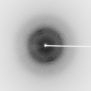
Dataset 107366_1_E1_###.mccd details
| Number of frames |
360 (1 - 360) |
| Distance [mm] |
200.0 |
| Oscillation width [°] |
0.25 |
| Phi [°] |
79.0 |
| Wavelength [Å] |
0.97905 |
| Experiment Date |
2009-02-18 |
| Equipment |
BL11-1
at SSRL (Stanford Synchrotron Radiation Laboratory)
|
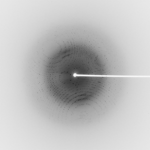
Dataset 107366_1_E2_###.mccd details
| Number of frames |
360 (1 - 360) |
| Distance [mm] |
200.0 |
| Oscillation width [°] |
0.25 |
| Phi [°] |
79.0 |
| Wavelength [Å] |
0.91837 |
| Experiment Date |
2009-02-18 |
| Equipment |
BL11-1
at SSRL (Stanford Synchrotron Radiation Laboratory)
|
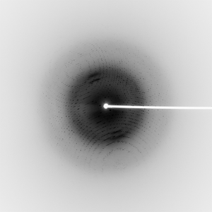
Dataset 107366_2_###.mccd details
| Number of frames |
360 (1 - 360) |
| Distance [mm] |
200.0 |
| Oscillation width [°] |
0.25 |
| Phi [°] |
79.0 |
| Wavelength [Å] |
0.97839 |
| Experiment Date |
2009-02-18 |
| Equipment |
BL11-1
at SSRL (Stanford Synchrotron Radiation Laboratory)
|
Dataset 107366_3_###.mccd details
| Number of frames |
360 (1 - 360) |
| Distance [mm] |
200.0 |
| Oscillation width [°] |
0.25 |
| Phi [°] |
79.0 |
| Wavelength [Å] |
0.91837 |
| Experiment Date |
2009-02-18 |
| Equipment |
BL11-1
at SSRL (Stanford Synchrotron Radiation Laboratory)
|