Diffraction project datasets FP9641B_3ihu
Project details
| Title |
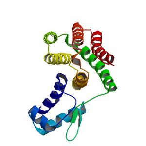
Crystal structure of DNA binding protein (YP_298823.1) from Ralstonia eutropha JMP134 at 1.92 A resolution |
| Authors |
Joint Center for Structural Genomics (JCSG) |
| Bioentity |
None |
| R / Rfree |
0.20 / 0.24 |
| Unit cell edges [Å] |
54.65 x
79.14 x
103.24
|
| Unit cell angles [°] |
90.0,
102.1,
90.0
|
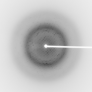
Dataset 116815_1_E1_###.mccd details
| Number of frames |
130 (1 - 130) |
| Distance [mm] |
250.0 |
| Oscillation width [°] |
1.00 |
| Phi [°] |
290.0 |
| Wavelength [Å] |
0.97862 |
| Experiment Date |
2009-04-16 |
| Equipment |
BL11-1
at SSRL (Stanford Synchrotron Radiation Laboratory)
|
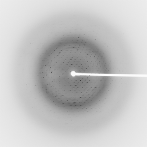
Dataset 116815_1_E2_###.mccd details
| Number of frames |
130 (1 - 130) |
| Distance [mm] |
250.0 |
| Oscillation width [°] |
1.00 |
| Phi [°] |
290.0 |
| Wavelength [Å] |
0.91837 |
| Experiment Date |
2009-04-16 |
| Equipment |
BL11-1
at SSRL (Stanford Synchrotron Radiation Laboratory)
|
Dataset 116815_2_###.mccd details
| Number of frames |
160 (1 - 160) |
| Distance [mm] |
250.0 |
| Oscillation width [°] |
1.00 |
| Phi [°] |
290.0 |
| Wavelength [Å] |
0.97799 |
| Experiment Date |
2009-04-16 |
| Equipment |
BL11-1
at SSRL (Stanford Synchrotron Radiation Laboratory)
|