Diffraction project datasets FP10366A_3di4
Project details
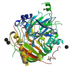
| Title |
Crystal structure of a duf1989 family protein (spo0365) from silicibacter pomeroyi dss-3 at 1.60 A resolution |
| Authors |
Joint Center for Structural Genomics (JCSG) |
| Bioentity |
None |
| R / Rfree |
0.14 / 0.18 |
| Unit cell edges [Å] |
51.47 x
95.76 x
58.69
|
| Unit cell angles [°] |
90.0,
109.0,
90.0
|
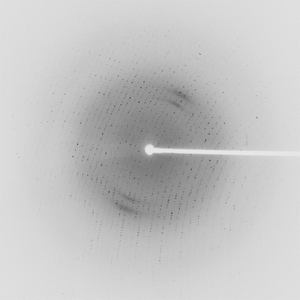
Dataset 85644_1_E1_###.mccd details
| Number of frames |
240 (1 - 240) |
| Distance [mm] |
250.0 |
| Oscillation width [°] |
0.50 |
| Phi [°] |
200.0 |
| Wavelength [Å] |
0.97941 |
| Experiment Date |
2008-05-13 |
| Equipment |
BL11-1
at SSRL (Stanford Synchrotron Radiation Laboratory)
|
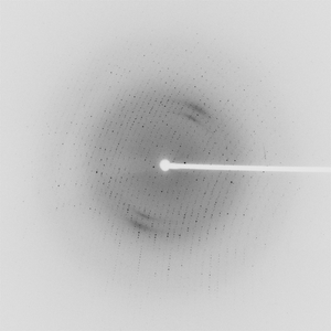
Dataset 85644_1_E2_###.mccd details
| Number of frames |
240 (1 - 240) |
| Distance [mm] |
250.0 |
| Oscillation width [°] |
0.50 |
| Phi [°] |
200.0 |
| Wavelength [Å] |
0.91837 |
| Experiment Date |
2008-05-13 |
| Equipment |
BL11-1
at SSRL (Stanford Synchrotron Radiation Laboratory)
|
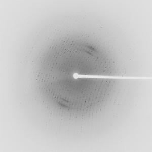
Dataset 85644_2_###.mccd details
| Number of frames |
240 (1 - 240) |
| Distance [mm] |
250.0 |
| Oscillation width [°] |
0.50 |
| Phi [°] |
200.0 |
| Wavelength [Å] |
0.97904 |
| Experiment Date |
2008-05-13 |
| Equipment |
BL11-1
at SSRL (Stanford Synchrotron Radiation Laboratory)
|