Diffraction project datasets FJ9298A_3pxp
- Method: SAD
- Resolution: 2.3 Å
- Space group: C 1 2 1
PDB website for 3PXP
PDB validation report for 3PXP
doi:10.18430/M33PXP
Project details
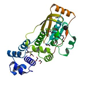
| Title | Crystal structure of a PAS and DNA binding domain containing protein (Caur_2278) from CHLOROFLEXUS AURANTIACUS J-10-FL at 2.30 A resolution |
| Authors | Xu, Q., van Wezel, G.P., Chiu, H.J., Jaroszewski, L., Klock, H.E., Knuth, M.W., Miller, M.D., Lesley, S.A., Godzik, A., Elsliger, M.A., Deacon, A.M., Wilson, I.A. |
| Bioentity | None |
| R / Rfree | 0.17 / 0.21 |
| Unit cell edges [Å] | 228.81 x 83.57 x 54.52 |
| Unit cell angles [°] | 90.0, 103.1, 90.0 |
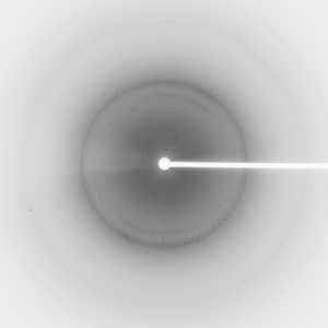
Dataset 44178_1_###.mccd details
| Number of frames | 350 (1 - 350) |
| Distance [mm] | 300.0 |
| Oscillation width [°] | 0.50 |
| Phi [°] | 152.0 |
| Wavelength [Å] | 0.97916 |
| Experiment Date | 2007-02-23 |
| Equipment | BL11-1 at SSRL (Stanford Synchrotron Radiation Laboratory) |