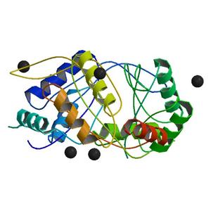
Diffraction project datasets 4iyq
- Method: Molecular Replacement
- Resolution: 2.55 Å
- Space group: P 1 21 1
PDB website for 4IYQ
PDB validation report for 4IYQ
doi:10.18430/M34IYQ
Project details
| Title | Crystal structure of divalent ion tolerance protein CutA1 from Ehrlichia chaffeensis |
| Authors | Seattle Structural Genomics Center for Infectious Disease (SSGCID), Abendroth, J., Sankaran, B., Buchko, G.W., Craig, T., Lorimer, D., Edwards, T.E. |
| R / Rfree | 0.17 / 0.22 |
| Unit cell edges [Å] | 87.49 x 32.58 x 89.08 |
| Unit cell angles [°] | 90.0, 119.7, 90.0 |
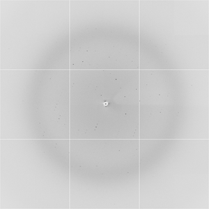
Dataset dcb18_15_1_###.img details
| Number of frames | 180 (1 - 180) |
| Distance [mm] | 370.0 |
| Oscillation width [°] | 1.00 |
| Omega [°] | 213.0 |
| Phi [°] | 213.0 |
| Wavelength [Å] | 0.97741 |
| Experiment Date | 2012-05-11 |
| Equipment | 5.0.1 at ALS (Advanced Light Source) |