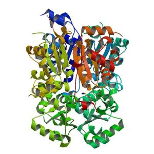
Diffraction project datasets 4f36
- Method: Molecular Replacement
- Resolution: 2.3 Å
- Space group: P 21 21 21
PDB website for 4F36
PDB validation report for 4F36
doi:10.18430/M34F36
Project details
| Title | Crystal structure of Nucleoside diphosphate kinase B from Trypanosoma brucei, apo form |
| Authors | Seattle Structural Genomics Center for Infectious Disease (SSGCID), Gardberg, A.S., Edwards, T.E., Staker, B., Stewart, L. |
| R / Rfree | 0.20 / 0.24 |
| Unit cell edges [Å] | 52.46 x 123.67 x 145.36 |
| Unit cell angles [°] | 90.0, 90.0, 90.0 |
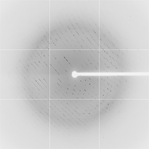
Dataset maz6-09_12_#####.img details
| Number of frames | 110 (1 - 110) |
| Distance [mm] | 340.0 |
| Oscillation width [°] | 1.00 |
| Omega [°] | 39.0 |
| Phi [°] | 39.0 |
| Wavelength [Å] | 1.03317 |
| Equipment | BL7-1 at SSRL (Stanford Synchrotron Radiation Laboratory) |