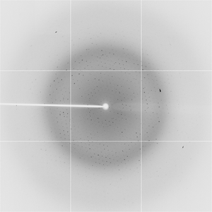
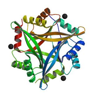
Diffraction project datasets 4e98


- Method: Molecular Replacement
- Resolution: 2.0 Å
- Space group: C 1 2 1
PDB website for 4E98
PDB validation report for 4E98
doi:10.18430/M34E98
Project details
| Title | Crystal structure of possible CutA1 divalent ion tolerance protein from Cryptosporidium parvum Iowa II |
| Authors | Buchko, G.W., Abendroth, J., Clifton, M.C., Robinson, H., Zhang, Y., Hewitt, S.N., Staker, B.L., Edwards, T.E., Van Voorhis, W.C., Myler, P.J. |
| R / Rfree | 0.20 / 0.25 |
| Unit cell edges [Å] | 94.46 x 55.59 x 67.29 |
| Unit cell angles [°] | 90.0, 108.2, 90.0 |
Dataset bd-5_5_###.img details

| Number of frames | 360 (1 - 360) |
| Distance [mm] | 250.0 |
| Oscillation width [°] | 1.00 |
| Wavelength [Å] | 1.07500 |
| Experiment Date | 2012-02-24 |
| Equipment | X29A at NSLS (National Synchrotron Light Source) |