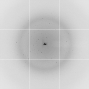
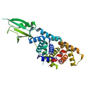
Diffraction project datasets 3v7o


- Method: Molecular Replacement
- Resolution: 2.25 Å
- Space group: P 21 21 21
PDB website for 3V7O
PDB validation report for 3V7O
doi:10.18430/M33V7O
Project details
| Title | Crystal structure of the C-terminal domain of Ebola virus VP30 (strain Reston-89) |
| Authors | Clifton, M.C., Kirchdoerfer, R.N., Atkins, K., Abendroth, J., Raymond, A., Grice, R., Barnes, S., Moen, S., Lorimer, D., Edwards, T.E., Myler, P.J., Saphire, E.O. |
| R / Rfree | 0.19 / 0.23 |
| Unit cell edges [Å] | 49.31 x 93.74 x 111.22 |
| Unit cell angles [°] | 90.0, 90.0, 90.0 |
Dataset fdi0-7_2_###.img details

| Number of frames | 240 (1 - 240) |
| Distance [mm] | 325.0 |
| Oscillation width [°] | 0.50 |
| Omega [°] | 180.0 |
| Phi [°] | 180.0 |
| Wavelength [Å] | 0.97740 |
| Experiment Date | 2011-12-09 |
| Equipment | 5.0.1 at ALS (Advanced Light Source) |