Diffraction project datasets 3uam
- Method: MR, MR
- Resolution: 2.0 Å
- Space group: P 1
PDB website for 3UAM
PDB validation report for 3UAM
doi:10.18430/M33UAM
Project details
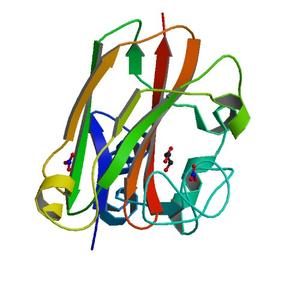
| Title | Crystal structure of a chitin binding domain from Burkholderia pseudomallei |
| Authors | Seattle Structural Genomics Center for Infectious Disease (SSGCID), Fox III, D., Gardberg, A., Armour, B., Staker, B., Stewart, L. |
| R / Rfree | 0.17 / 0.21 |
| Unit cell edges [Å] | 70.92 x 74.78 x 74.77 |
| Unit cell angles [°] | 120.0, 98.0, 103.0 |
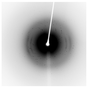
Dataset 225965f8_x####.img details
| Number of frames | 720 (1 - 720) |
| Distance [mm] | 50.0 |
| Oscillation width [°] | 0.50 |
| Omega [°] | -95.0 |
| Wavelength [Å] | 1.54178 |
| Equipment | HOME_SOURCE at HOME_SOURCE (Home Source) |
Dataset 225965f8_y####.img details
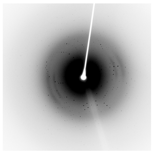
| Number of frames | 720 (1 - 720) |
| Distance [mm] | 50.0 |
| Oscillation width [°] | 0.50 |
| Omega [°] | -95.0 |
| Kappa / Chi [°] | 15.0 |
| Wavelength [Å] | 1.54178 |
| Equipment | HOME_SOURCE at HOME_SOURCE (Home Source) |