Diffraction project datasets 3sx2
- Method: MR
- Resolution: 1.5 Å
- Space group: P 1
PDB website for 3SX2
PDB validation report for 3SX2
doi:10.18430/M33SX2
Project details
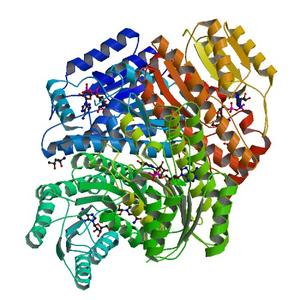
| Title | Crystal structure of a putative 3-ketoacyl-(acyl-carrier-protein) reductase from Mycobacterium paratuberculosis in complex with NAD |
| Authors | Baugh, L., Phan, I., Begley, D.W., Clifton, M.C., Armour, B., Dranow, D.M., Taylor, B.M., Muruthi, M.M., Abendroth, J., Fairman, J.W., Fox, D., Dieterich, S.H., Staker, B.L., Gardberg, A.S., Choi, R., Hewitt, S.N., Napuli, A.J., Myers, J., Barrett, L.K., Zhang, Y., Ferrell, M., Mundt, E., Thompkins, K., Tran, N., Lyons-Abbott, S., Abramov, A., Sekar, A., Serbzhinskiy, D., Lorimer, D., Buchko, G.W., Stacy, R., Stewart, L.J., Edwards, T.E., Van Voorhis, W.C., Myler, P.J. |
| R / Rfree | 0.14 / 0.16 |
| Unit cell edges [Å] | 64.46 x 70.76 x 125.68 |
| Unit cell angles [°] | 97.0, 93.9, 86.9 |
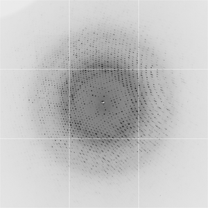
Dataset yup6-2_13_###.img details
| Number of frames | 360 (1 - 360) |
| Distance [mm] | 200.0 |
| Oscillation width [°] | 1.00 |
| Wavelength [Å] | 0.97650 |
| Experiment Date | 2010-12-21 |
| Equipment | 5.0.3 at ALS (Advanced Light Source) |