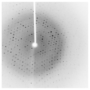
Diffraction project datasets 3sjs
- Method: Molecular Replacement
- Resolution: 1.9 Å
- Space group: C 2 2 21
PDB website for 3SJS
PDB validation report for 3SJS
doi:10.18430/M33SJS
Project details
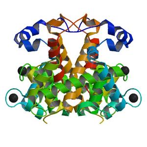
| Title | Crystal structure of URE3-binding protein from Entamoeba histolytica, (D127A,N129A) mutant, native form |
| Authors | Seattle Structural Genomics Center for Infectious Disease (SSGCID), Gardberg, A., Edwards, T., Staker, B., Skubak, P., Gilchrist, C., Stewart, L. |
| R / Rfree | 0.17 / 0.22 |
| Unit cell edges [Å] | 46.25 x 68.29 x 130.89 |
| Unit cell angles [°] | 90.0, 90.0, 90.0 |
Dataset native-ph-4.6-x####.img details
| Number of frames | 417 (1 - 417) |
| Distance [mm] | 60.0 |
| Oscillation width [°] | 0.50 |
| Omega [°] | -80.0 |
| Wavelength [Å] | 1.54178 |
| Equipment | HOME_SOURCE at HOME_SOURCE (Home Source) |