Diffraction project datasets 3sdw
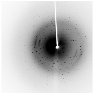
- Method: Molecular Replacement
- Resolution: 1.8 Å
- Space group: F 2 2 2
PDB website for 3SDW
PDB validation report for 3SDW
doi:10.18430/M33SDW
Project details
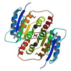
| Title | Crystal structure of a ribose-5-phosphate isomerase B RpiB from Coccidioides immitis bound to phosphate |
| Authors | Edwards, T.E., Abramov, A.B., Smith, E.R., Baydo, R.O., Leonard, J.T., Leibly, D.J., Thompkins, K.B., Clifton, M.C., Gardberg, A.S., Staker, B.L., Van Voorhis, W.C., Myler, P.J., Stewart, L.J. |
| R / Rfree | 0.15 / 0.18 |
| Unit cell edges [Å] | 77.70 x 85.22 x 96.29 |
| Unit cell angles [°] | 90.0, 90.0, 90.0 |
Dataset 222975a2_x####.img details
| Number of frames | 340 (1 - 340) |
| Distance [mm] | 50.0 |
| Oscillation width [°] | 1.00 |
| Omega [°] | -90.0 |
| Wavelength [Å] | 1.54178 |
| Equipment | HOME_SOURCE at HOME_SOURCE (Home Source) |