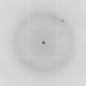
Diffraction project datasets 3sbx
- Method: Molecular Replacement
- Resolution: 2.5 Å
- Space group: P 1
PDB website for 3SBX
PDB validation report for 3SBX
doi:10.18430/M33SBX
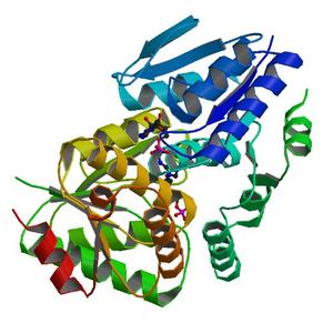
Project details
| Title | Crystal structure of a putative uncharacterized protein from Mycobacterium marinum bound to adenosine 5'-monophosphate AMP |
| Authors | Baugh, L., Phan, I., Begley, D.W., Clifton, M.C., Armour, B., Dranow, D.M., Taylor, B.M., Muruthi, M.M., Abendroth, J., Fairman, J.W., Fox, D., Dieterich, S.H., Staker, B.L., Gardberg, A.S., Choi, R., Hewitt, S.N., Napuli, A.J., Myers, J., Barrett, L.K., Zhang, Y., Ferrell, M., Mundt, E., Thompkins, K., Tran, N., Lyons-Abbott, S., Abramov, A., Sekar, A., Serbzhinskiy, D., Lorimer, D., Buchko, G.W., Stacy, R., Stewart, L.J., Edwards, T.E., Van Voorhis, W.C., Myler, P.J. |
| R / Rfree | 0.24 / 0.29 |
| Unit cell edges [Å] | 67.24 x 80.12 x 85.69 |
| Unit cell angles [°] | 106.7, 90.1, 98.8 |
Dataset djk5-15_1_###.img details
| Number of frames | 315 (1 - 315) |
| Distance [mm] | 365.0 |
| Oscillation width [°] | 1.00 |
| Omega [°] | -35.0 |
| Phi [°] | -35.0 |
| Wavelength [Å] | 0.97740 |
| Experiment Date | 2011-04-11 |
| Equipment | 5.0.1 at ALS (Advanced Light Source) |