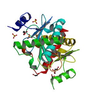
Diffraction project datasets 3p0t
- Method: Molecular Replacement
- Resolution: 1.9 Å
- Space group: P 43 21 2
PDB website for 3P0T
PDB validation report for 3P0T
doi:10.18430/M33P0T
Project details
| Title | Crystal structure of an HIT-like protein from mycobacterium paratuberculosis |
| Authors | Baugh, L., Phan, I., Begley, D.W., Clifton, M.C., Armour, B., Dranow, D.M., Taylor, B.M., Muruthi, M.M., Abendroth, J., Fairman, J.W., Fox, D., Dieterich, S.H., Staker, B.L., Gardberg, A.S., Choi, R., Hewitt, S.N., Napuli, A.J., Myers, J., Barrett, L.K., Zhang, Y., Ferrell, M., Mundt, E., Thompkins, K., Tran, N., Lyons-Abbott, S., Abramov, A., Sekar, A., Serbzhinskiy, D., Lorimer, D., Buchko, G.W., Stacy, R., Stewart, L.J., Edwards, T.E., Van Voorhis, W.C., Myler, P.J. |
| R / Rfree | 0.16 / 0.21 |
| Unit cell edges [Å] | 55.61 x 55.61 x 205.28 |
| Unit cell angles [°] | 90.0, 90.0, 90.0 |
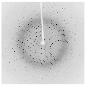
Dataset 217599g11_x####.img details
| Number of frames | 675 (1 - 675) |
| Distance [mm] | 50.0 |
| Oscillation width [°] | 0.20 |
| Omega [°] | -45.0 |
| Wavelength [Å] | 1.54178 |
| Equipment | HOME_SOURCE at HOME_SOURCE (Home Source) |