Diffraction project datasets 3mxu

- Method: Molecular Replacement
- Resolution: 1.8 Å
- Space group: H 3 2
PDB website for 3MXU
PDB validation report for 3MXU
doi:10.18430/M33MXU
Project details
| Title | Crystal structure of glycine cleavage system protein H from Bartonella henselae |
| Authors | Edwards, T.E., Gardberg, A.S., Seattle Structural Genomics Center for Infectious Disease (SSGCID) |
| R / Rfree | 0.18 / 0.20 |
| Unit cell edges [Å] | 98.91 x 98.91 x 131.54 |
| Unit cell angles [°] | 90.0, 90.0, 120.0 |
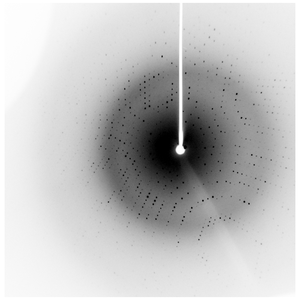
Dataset 206762b1_a####.img details
| Number of frames | 160 (1 - 160) |
| Distance [mm] | 50.0 |
| Oscillation width [°] | 1.00 |
| Omega [°] | -100.0 |
| Kappa / Chi [°] | 30.0 |
| Wavelength [Å] | 1.54178 |
| Equipment | HOME_SOURCE at HOME_SOURCE (Home Source) |