Diffraction project datasets 3mx6
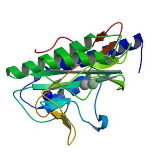
- Method: Molecular Replacement
- Resolution: 1.7 Å
- Space group: P 1 21 1
PDB website for 3MX6
PDB validation report for 3MX6
doi:10.18430/M33MX6
Project details
| Title | Crystal structure of methionine aminopeptidase from Rickettsia prowazekii bound to methionine |
| Authors | Helgren, T.R., Chen, C., Wangtrakuldee, P., Edwards, T.E., Staker, B.L., Abendroth, J., Sankaran, B., Housley, N.A., Myler, P.J., Audia, J.P., Horn, J.R., Hagen, T.J. |
| R / Rfree | 0.17 / 0.21 |
| Unit cell edges [Å] | 50.37 x 67.55 x 80.89 |
| Unit cell angles [°] | 90.0, 97.4, 90.0 |
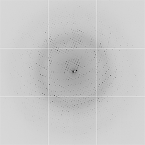
Dataset jkz1_10_1_###.img details
| Number of frames | 249 (1 - 249) |
| Distance [mm] | 260.0 |
| Oscillation width [°] | 1.00 |
| Wavelength [Å] | 0.97740 |
| Experiment Date | 2010-04-30 |
| Equipment | 5.0.1 at ALS (Advanced Light Source) |