Diffraction project datasets 3ift
- Method: Molecular Replacement
- Resolution: 2.0 Å
- Space group: C 1 2 1
PDB website for 3IFT
PDB validation report for 3IFT
doi:10.18430/M33IFT
Project details
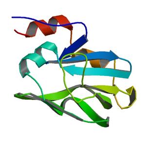
| Title | Crystal structure of glycine cleavage system protein H from Mycobacterium tuberculosis, using X-rays from the Compact Light Source. |
| Authors | Abendroth, J., McCormick, M.S., Edwards, T.E., Staker, B., Loewen, R., Gifford, M., Rifkin, J., Mayer, C., Guo, W., Zhang, Y., Myler, P., Kelley, A., Analau, E., Hewitt, S.N., Napuli, A.J., Kuhn, P., Ruth, R.D., Stewart, L.J. |
| R / Rfree | 0.18 / 0.25 |
| Unit cell edges [Å] | 86.45 x 51.01 x 32.57 |
| Unit cell angles [°] | 90.0, 95.1, 90.0 |
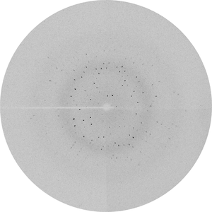
Dataset decode-LTI_209b-position3_newcryo_07-15-200903.#### details
| Number of frames | 70 (1 - 70) |
| Distance [mm] | 120.0 |
| Oscillation width [°] | 1.00 |
| Phi [°] | 200.0 |
| Wavelength [Å] | 0.81836 |
| Experiment Date | 2009-07-15 |
| Equipment | HOME_SOURCE at HOME_SOURCE (Home Source) |
Dataset decode-LTI_209b-position3_newcryo_07-15-200905.#### details
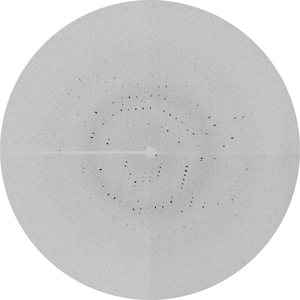
| Number of frames | 45 (2 - 46) |
| Distance [mm] | 120.0 |
| Oscillation width [°] | 1.00 |
| Phi [°] | 156.0 |
| Wavelength [Å] | 0.81836 |
| Experiment Date | 2009-07-20 |
| Equipment | HOME_SOURCE at HOME_SOURCE (Home Source) |