Diffraction project datasets 3he2
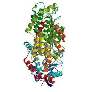
- Method: MR, MR
- Resolution: 2.3 Å
- Space group: P 21 21 21
PDB website for 3HE2
PDB validation report for 3HE2
doi:10.18430/M33HE2
Project details
| Title | Crystal structure of enoyl-CoA hydratase from Mycobacterium tuberculosis |
| Authors | Baugh, L., Phan, I., Begley, D.W., Clifton, M.C., Armour, B., Dranow, D.M., Taylor, B.M., Muruthi, M.M., Abendroth, J., Fairman, J.W., Fox, D., Dieterich, S.H., Staker, B.L., Gardberg, A.S., Choi, R., Hewitt, S.N., Napuli, A.J., Myers, J., Barrett, L.K., Zhang, Y., Ferrell, M., Mundt, E., Thompkins, K., Tran, N., Lyons-Abbott, S., Abramov, A., Sekar, A., Serbzhinskiy, D., Lorimer, D., Buchko, G.W., Stacy, R., Stewart, L.J., Edwards, T.E., Van Voorhis, W.C., Myler, P.J. |
| R / Rfree | 0.19 / 0.24 |
| Unit cell edges [Å] | 70.12 x 76.47 x 134.27 |
| Unit cell angles [°] | 90.0, 90.0, 90.0 |
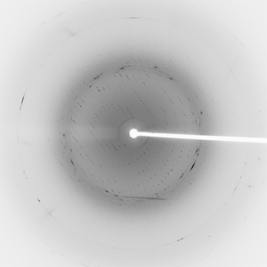
Dataset puck-031-16_16_###.mccd details
| Number of frames | 120 (1 - 120) |
| Distance [mm] | 300.0 |
| Oscillation width [°] | 1.00 |
| Wavelength [Å] | 0.97946 |
| Experiment Date | 2009-04-04 |
| Equipment | BL12-2 at SSRL (Stanford Synchrotron Radiation Laboratory) |