Diffraction project datasets 3gvg
- Method: MR
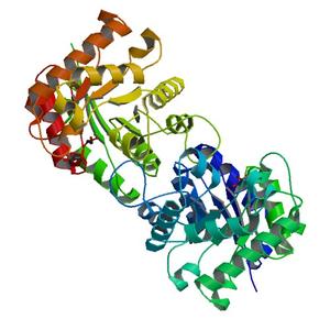
- Resolution: 1.55 Å
- Space group: C 1 2 1
PDB website for 3GVG
PDB validation report for 3GVG
doi:10.18430/M33GVG
Project details
| Title | Crystal structure of Triosephosphate isomerase from Mycobacterium tuberculosis |
| Authors | Baugh, L., Phan, I., Begley, D.W., Clifton, M.C., Armour, B., Dranow, D.M., Taylor, B.M., Muruthi, M.M., Abendroth, J., Fairman, J.W., Fox, D., Dieterich, S.H., Staker, B.L., Gardberg, A.S., Choi, R., Hewitt, S.N., Napuli, A.J., Myers, J., Barrett, L.K., Zhang, Y., Ferrell, M., Mundt, E., Thompkins, K., Tran, N., Lyons-Abbott, S., Abramov, A., Sekar, A., Serbzhinskiy, D., Lorimer, D., Buchko, G.W., Stacy, R., Stewart, L.J., Edwards, T.E., Van Voorhis, W.C., Myler, P.J. |
| R / Rfree | 0.14 / 0.17 |
| Unit cell edges [Å] | 133.12 x 55.76 x 76.42 |
| Unit cell angles [°] | 90.0, 104.0, 90.0 |
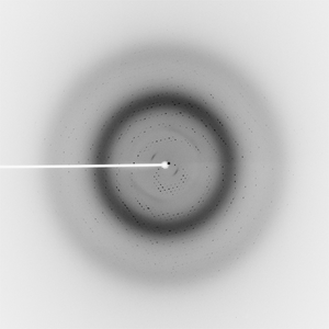
Dataset cps041-11_1.#### details
| Number of frames | 180 (1 - 180) |
| Distance [mm] | 200.0 |
| Oscillation width [°] | 1.00 |
| Phi [°] | -120.0 |
| Wavelength [Å] | 0.97933 |
| Experiment Date | 2009-03-20 |
| Equipment | 23-ID-D at APS (Advanced Photon Source) |