Diffraction project datasets 2636534_1vrb
Project details
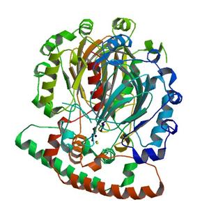
| Title |
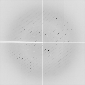
Crystal structure of Putative asparaginyl hydroxylase (2636534) from Bacillus subtilis at 2.60 A resolution |
| Authors |
Joint Center for Structural Genomics (JCSG) |
| Bioentity |
None |
| R / Rfree |
0.22 / 0.27 |
| Unit cell edges [Å] |
47.30 x
104.66 x
286.61
|
| Unit cell angles [°] |
90.0,
90.0,
90.0
|
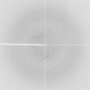
Dataset T1570_1_####.img details
| Number of frames |
280 (1000 - 1279) |
| Distance [mm] |
261.0 |
| Oscillation width [°] |
0.10 |
| Phi [°] |
125.4 |
| Wavelength [Å] |
1.03320 |
| Experiment Date |
2003-09-22 |
| Equipment |
8.3.1
at ALS (Advanced Light Source)
|
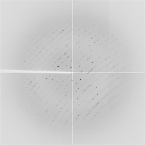
Dataset T1570_1_####.img details
| Number of frames |
220 (1281 - 1500) |
| Distance [mm] |
261.0 |
| Oscillation width [°] |
0.10 |
| Phi [°] |
153.4 |
| Wavelength [Å] |
1.03320 |
| Experiment Date |
2003-09-22 |
| Equipment |
8.3.1
at ALS (Advanced Light Source)
|
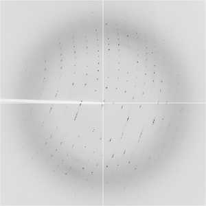
Dataset T1570_1_###.img details
| Number of frames |
552 (4 - 555) |
| Distance [mm] |
261.0 |
| Oscillation width [°] |
0.10 |
| Phi [°] |
26.2 |
| Wavelength [Å] |
1.03320 |
| Experiment Date |
2003-09-22 |
| Equipment |
8.3.1
at ALS (Advanced Light Source)
|
Dataset T1570_1_###.img details
| Number of frames |
443 (557 - 999) |
| Distance [mm] |
261.0 |
| Oscillation width [°] |
0.10 |
| Phi [°] |
81.0 |
| Wavelength [Å] |
1.03320 |
| Experiment Date |
2003-09-22 |
| Equipment |
8.3.1
at ALS (Advanced Light Source)
|