Diffraction project datasets 16741099_1vm8
Project details
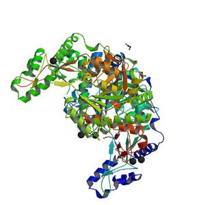
| Title |
Crystal structure of UDP-N-acetylglucosamine pyrophosphorylase (Agx2) from Mus musculus at 2.50 A resolution |
| Authors |
Joint Center for Structural Genomics (JCSG) |
| Bioentity |
None |
| R / Rfree |
0.21 / 0.27 |
| Unit cell edges [Å] |
80.09 x
73.40 x
107.13
|
| Unit cell angles [°] |
90.0,
99.8,
90.0
|
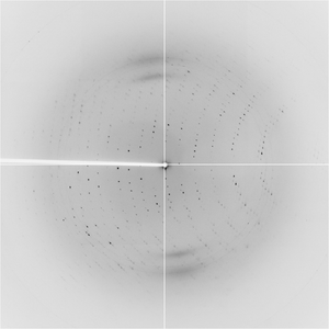
Dataset 3985_1_###.img details
| Number of frames |
108 (1 - 108) |
| Distance [mm] |
250.0 |
| Oscillation width [°] |
1.50 |
| Phi [°] |
77.0 |
| Wavelength [Å] |
0.97920 |
| Experiment Date |
2003-06-05 |
| Equipment |
8.2.1
at ALS (Advanced Light Source)
|
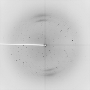
Dataset 3985_2_###.img details
| Number of frames |
108 (1 - 108) |
| Distance [mm] |
250.0 |
| Oscillation width [°] |
1.50 |
| Phi [°] |
77.0 |
| Wavelength [Å] |
1.00000 |
| Experiment Date |
2003-06-05 |
| Equipment |
8.2.1
at ALS (Advanced Light Source)
|
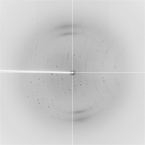
Dataset 3985_4_###.img details
| Number of frames |
108 (1 - 108) |
| Distance [mm] |
250.0 |
| Oscillation width [°] |
1.50 |
| Phi [°] |
77.0 |
| Wavelength [Å] |
0.97910 |
| Experiment Date |
2003-06-05 |
| Equipment |
8.2.1
at ALS (Advanced Light Source)
|