Diffraction project datasets 13421216_1vjf
Project details
| Title |
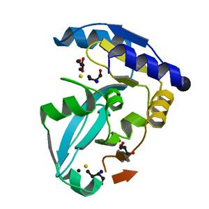
CRYSTAL STRUCTURE OF A PUTATIVE DNA-BINDING PROTEIN (CC_0111) FROM CAULOBACTER CRESCENTUS CB15 AT 1.62 A RESOLUTION |
| Authors |
Joint Center for Structural Genomics (JCSG) |
| Bioentity |
None |
| R / Rfree |
0.15 / 0.17 |
| Unit cell edges [Å] |
50.89 x
50.89 x
121.43
|
| Unit cell angles [°] |
90.0,
90.0,
90.0
|
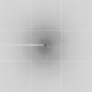
Dataset T1823_1_E1_###.img details
| Number of frames |
90 (1 - 90) |
| Distance [mm] |
290.0 |
| Oscillation width [°] |
1.00 |
| Phi [°] |
351.0 |
| Wavelength [Å] |
0.97960 |
| Experiment Date |
2003-10-01 |
| Equipment |
8.2.2
at ALS (Advanced Light Source)
|
Dataset T1823_1_E2_###.img details
| Number of frames |
90 (1 - 90) |
| Distance [mm] |
290.0 |
| Oscillation width [°] |
1.00 |
| Phi [°] |
351.0 |
| Wavelength [Å] |
1.00000 |
| Experiment Date |
2003-10-01 |
| Equipment |
8.2.2
at ALS (Advanced Light Source)
|
Dataset T1823_2_###.img details
| Number of frames |
90 (1 - 90) |
| Distance [mm] |
290.0 |
| Oscillation width [°] |
1.00 |
| Phi [°] |
351.0 |
| Wavelength [Å] |
0.97940 |
| Experiment Date |
2003-10-01 |
| Equipment |
8.2.2
at ALS (Advanced Light Source)
|
Dataset T1823_4_###.img details
| Number of frames |
135 (1 - 135) |
| Distance [mm] |
204.0 |
| Oscillation width [°] |
0.75 |
| Phi [°] |
81.01 |
| Wavelength [Å] |
1.00000 |
| Experiment Date |
2003-10-01 |
| Equipment |
8.2.2
at ALS (Advanced Light Source)
|